Mammography
Schedule your mammogram today
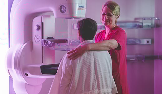
Your First Line of Defense
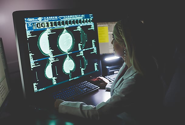
For breast cancer screening and investigation of symptoms, mammography is usually the best place to start. NMHS offers the most advanced technology, 3D mammography, in seven locations.
Mammograms use X-rays to examine breast tissues. 3D mammography units take multiple X-rays to create a 3D image of the breast. Mammograms can be used in two ways:
Screening, where the patient has no symptoms or problems but is at risk
Diagnostic, which investigates symptoms or follow up on results from other exams
One in eight women will be diagnosed with breast cancer in their lifetime. Breast cancer is the second leading cause of cancer death in U.S. women. Screening mammograms reduce the risk of dying from breast cancer for women between the ages of 40 and 74 and can find breast cancer before symptoms appear. Most insurance covers screening mammograms with no out-of-pocket cost.
The American College of Radiology recommends:
Annual mammograms starting at age 40 for those of average risk
Early assessment and increased screening for those at high risk
Even if you are not yet 40 years old or it’s been less than a year since your last screening mammogram, there are breast symptoms that should be evaluated. If you are experiencing any of these symptoms, please contact your healthcare provider so that the appropriate exam may be ordered. National guidelines recommend diagnostic imaging for any of these conditions:
Breast lump
Breast pain or tenderness
Nipple discharge
Nipple retraction
As radiologists examine the mammogram images, they are looking certain diagnostic markers in the breast tissue. If the radiologist sees an area that is questionable, unclear, abnormal or varies from your previous film, you will be asked to return for additional views or further evaluation of the area. This does not mean that you have cancer; rather, it means that the radiologist needs additional information to be more confident that the suspicious area is clearly a normal finding. The use of 3D mammography has reduced the need for many callbacks. Diagnostic markers include:
Microcalcifications
Masses
Asymmetries
Distortions
Breast tissue density
Changes from previous mammograms
A key diagnostic marker that radiologists look for on mammograms is microcalificiations. By looking at the size, shape and distribution of the microcalcifications, the radiologist can detect whether these microcalcifications may indicate a possible malignancy.
Most microcalcifications are benign; only a small percentage indicate cancer.
Normal metabolism can cause microcalcifications; not related to dietary intake.
In some cases, they can be the first sign of a small breast cancer.
It can be difficult to differentiate the benign from the suspicious.
Next steps can include short-term follow up, surgical consult or other tests.
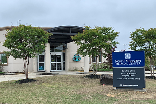
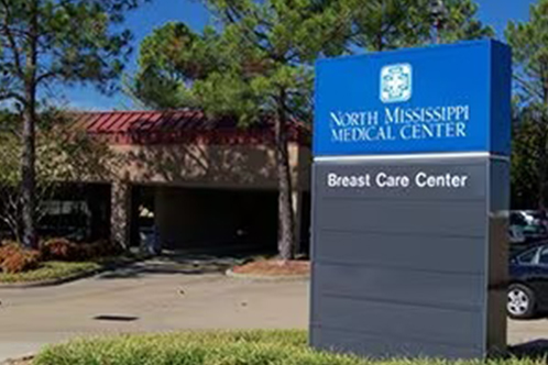
Related Locations


Upholding high standards for comprehensive breast care

ACS accredits NMMC’s breast care program
Meets national standards for comprehensive, patient-centered, multidisciplinary care.

ACR Designated Comprehensive Breast Imaging Center
NMMC Breast Care Center designated comprehensive center by American College of Radiology
Mammography
Accredited by the American College of Radiology in mammography
Breast Ultrasound
Accredited by the American College of Radiology in breast ultrasound
Stereotactic Breast Biopsy
Accredited by the American College of Radiology in stereotactic breast biopsy
Magnetic Resonance Imaging (MRI)
Accredited in MRI from American College of Radiology includes breast MRI
Patient Stories

Mammograms Are Not Optional
Paige McFall was determined to beat her aggressive breast cancer.


‘I Thought What I Found Would Go Away, But it Didn’t'
Like many busy women, Debbie Cochran put off getting her annual mammogram.

‘I Was Fortunate to Catch it as Early as We Did’
If Anita Monroe had put off her mammogram, her prognosis would not be so positive.

‘If Something Is Not Right, Get Checked’
Shirlette Judon celebrating 16 years as breast cancer survivor
Mammograms Are Not Optional
Paige McFall was determined to beat her aggressive breast cancer.

‘I Thought What I Found Would Go Away, But it Didn’t'
Like many busy women, Debbie Cochran put off getting her annual mammogram.
‘I Was Fortunate to Catch it as Early as We Did’
If Anita Monroe had put off her mammogram, her prognosis would not be so positive.
‘If Something Is Not Right, Get Checked’
Shirlette Judon celebrating 16 years as breast cancer survivor
Related Resources
View AllBreast cancer screening matters. Annual screening with mammography is recommended starting at age 40 for those at average risk.

Breast Density
Breast density impacts the effectiveness for mammography and the risk of breast cancer. Supplemental screening with automated ultrasound (ABUS) along with mammography improves breast cancer detection rate.
Breast Density
Breast density impacts the effectiveness for mammography and the risk of breast cancer. Supplemental screening with automated ultrasound (ABUS) along with mammography improves breast cancer detection rate.