Have a doctor’s order for an imaging study?
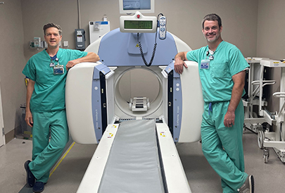
Powerful imaging technology
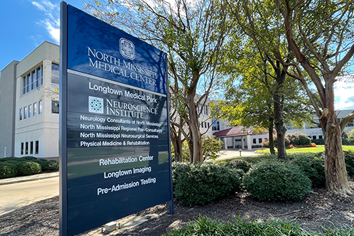
Nuclear medicine, which includes PET scans, may sound intimidating, but it’s as safe as X-ray. Medical imaging using nuclear medicine is available at Longtown Medical Imaging in Tupelo.
Nuclear medicine is a branch of medical imaging that uses small amounts of radioactive material to diagnose and determine the severity and/or treat a variety of diseases. Nuclear imaging findings may even eliminate the need for exploratory surgery. Some common uses for nuclear medicine include:
Observing the progression of heart disease
Detecting disorders in bones, gall bladder disease & intestinal bleeding
Tracking metabolic activity
Diagnosis of Parkinson’s disease
Where CT scans and MRIs are designed to look at the structure of body parts, positron emission tomography, or PET, measures the metabolic activity of body tissues. During a PET scan, a radioactive substance called a tracer is combined with a chemical substance and either inhaled or injected into a vein. The tracer emits positrons—tiny, positively charged particles that produce signals. A special camera records the tracer's signals as it travels through the body and collects in organs. A computer then converts the signals into three-dimensional images of the examined organ. PET scans can:
Analyze cancer tumors to track progression, response to treatment & possible spread
Stage cancers, using a combination PET/CT scanner
Examine brain function
Distinguish Alzheimer’s disease from other forms of dementia
Evaluate heart metabolism & blood flow
Take between 30 minutes & three hours to perform
Related Locations
Imaging Awards
Related Resources
View AllNurse Link® is a free telephone triage and health information service provided by North Mississippi Health Services. Using computerized medical protocols, nurses direct callers to the most appropriate medical treatment.

Cancer Screening
Radiology plays an important part in screening and early detection for breast cancer and lung cancer. Breast cancer screening starts with mammography. Lung cancer screening uses low dose CT.

Cancer Screening
Radiology plays an important part in screening and early detection for breast cancer and lung cancer. Breast cancer screening starts with mammography. Lung cancer screening uses low dose CT.

