Ultrasound & Vascular Studies
Have a doctor’s order for an imaging study?
Sound Into Images
Ultrasound imaging, also called sonography, is an imaging method that uses high-frequency sound waves to produce images of structures within your body.
Ultrasound images can provide valuable information for diagnosing and treating a variety of diseases and conditions. Because ultrasound imaging uses sound waves instead of radiation, it is very safe and can be used in several ways. Ultrasound images are captured in real-time.
Ultrasound is available at all NMHS hospitals and outpatient imaging clinics. Common uses for ultrasound include:
Monitoring pregnant women & their unborn infants
Checking the structure of internal organs
Tracking movement inside the body, such as the movement of blood through the heart & blood vessels
Guiding minimally invasive procedures like biopsies or removal of fluid
Ultrasound technology is used for vascular studies as well, which can help diagnose peripheral artery disease (PAD) and atherosclerosis.
Atherosclerosis causes a buildup of plaque in the blood vessels.
Atherosclerosis affects many blood vessels in the body.
PAD occurs when the plaque reduces or stops blood flow to the extremities.
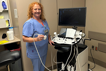
During an ultrasound exam, sound waves pass through the body. The ultrasound machine detects the echoes are produced by the sound waves. Those echoes help identify the size, shape and density of tissues inside the body. During an ultrasound exam:
An ultrasound technologist will place a transducer against your skin for the exam.
The transducer sends & receives the sound waves.
The technologist will apply a liquid gel to the skin.
The gel helps ensures sound waves are freely conducted.
The technologist sweeps the transducer back & forth over the area of interest.
The echoes are instantly measured & displayed by a computer.
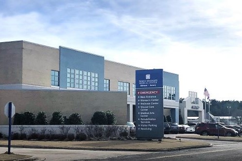
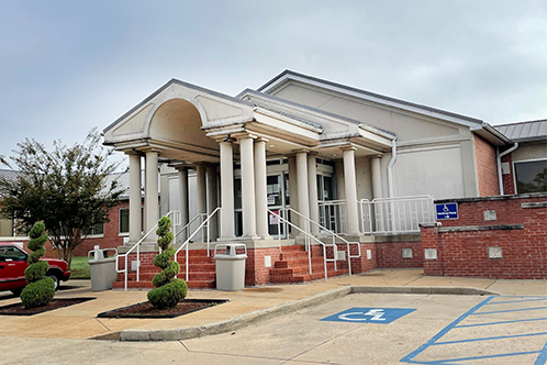
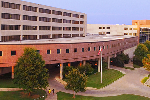
Related Locations


Imaging Awards
Mammography
Accredited by the American College of Radiology
Computed Tomography (CT)
Accredited by the American College of Radiology
Magnetic Resonance Imaging (MRI)
Accredited by the American College of Radiology
Radiology Department
Accredited by the State of Mississippi
Related Resources
View AllNurse Link® is a free telephone triage and health information service provided by North Mississippi Health Services. Using computerized medical protocols, nurses direct callers to the most appropriate medical treatment.
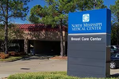
Cancer Screening
Radiology plays an important part in screening and early detection for breast cancer and lung cancer. Breast cancer screening starts with mammography. Lung cancer screening uses low dose CT.
Cancer Screening
Radiology plays an important part in screening and early detection for breast cancer and lung cancer. Breast cancer screening starts with mammography. Lung cancer screening uses low dose CT.