- Medical Services
- Heart and Vascular Care
- Conditions We Treat
- Atrial Fibrillation

Atrial Fibrillation
At North Mississippi Medical Center, you will find the most advanced treatment options for atrial fibrillation.
Your Heart is in the Right Place
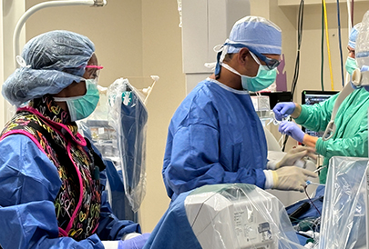
NMMC electrophysiologists (cardiologists who specialize in heart rhythm conditions) collaborate with cardiothoracic surgeons for the most appropriate treatment for atrial fibrillation.
Atrial fibrillation is an irregular heart rhythm that originates in the atria (top chambers of the heart). Instead of the electrical impulse traveling in an orderly fashion through the heart, many impulses begin and spread through the atria causing the atria to quiver or fibrillate. Some of the electrical impulses travel through the heart and make the ventricles (bottom chambers of the heart) squeeze or contract.
Consequences
Atrial fibrillation is not life threatening, but it can be extremely bothersome and sometime dangerous. People with atrial fibrillation are seven times more likely to have a stroke. Blood clots can travel to other parts of the body (heart, lungs, kidneys, intestines), causing damage. If your heart rate is fast over a long period of time, it can cause heart failure. Chronic atrial fibrillation can increase your risk of death.
Atrial fibrillation episodes may last minutes, hours or days. You may have atrial fibrillation without having any symptoms. But, if symptoms do occur, they may include:
Heart palpitations (skipping or racing sensation in your chest)
Shortness of breath (difficulty breathing during normal activities, climbing stairs or walking long distances)
Dizziness (lightheadedness or feeling faint)
Weakness (lack of energy)
Discomfort, pain or pressure in chest
Several different conditions can cause atrial fibrillation. Your risk increases with age, especially after age 60. Here are some of the most common causes of atrial fibrillation.
Hypertension (high blood pressure)
Heart failure
Cardiomyopathy (heart enlargement)
Coronary artery disease
Heart valve diseas
Previous heart surgery
Congenital heart disease
Pulmonary embolism
Thyroid problems
Treatment
Treatment for atrial fibrillation includes restoring and maintaining a normal heart rhythm, controlling the heart rate and preventing blood clots to reduce the risk of a stroke. Medications, lifestyle changes, procedures and surgery treat atrial fibrillation.
Medications
- Rhythm control medications help restore or maintain a normal heart rhythm (sinus rhythm). You may need to stay in the hospital when you first start taking these medications so we can monitor your heart rhythm and response to the medication. You may need these medications indefinitely. Unfortunately, they may lose their effectiveness over time.
- Rate control medications slow the heart rate and do not control the heart rhythm.
- Medications to prevent blood clots reduce (but do not eliminate) your risk of stroke. Anticoagulant or antiplatelet therapy medications, such as warfarin (Coumadin) are common. Depending on your medical history, aspirin may be used instead of warfarin.
Lifestyle Changes
- Limit or avoid caffeine and other stimulants (such as coffee, tea, and some over-the-counter medications).
- Limit your intake of alcohol.
- Stop smoking.
Procedures & Surgery
- Electrical cardioversion: We give a short-acting anesthesia or sedative then deliver an electrical shock to your heart through patches or paddles placed on your chest. This procedure frequently restores a normal rhythm, but its effect may not be permanent.
- Electrophysiology study: This procedure allows doctors to determine exactly what your rhythm problem is and choose appropriate treatment.
- Catheter ablation: We use ablation therapy for people who cannot tolerate medications or when medications cannot maintain a normal heart rhythm. A cardiac electrophysiologist uses various energy sources (radiofrequency and cryo) to destroy (ablate) abnormal heart tissue that causes or sustains the abnormal heart rhythm. Cardiac ablation treats atrial fibrillation, atrial flutter, supraventricular tachycardia, Wolff-Parkinson-White (WPW) syndrome and ventricular tachycardia. We perform pulmonary vein isolation or ablation of the AV node:
- Pulmonary vein isolation: Research has proven that most atrial fibrillation signals come from the four pulmonary veins. During this procedure, we insert special catheters into the blood vessels of the atrium. We use a catheter to locate the abnormal impulses coming from the pulmonary veins. Another catheter delivers the radiofrequency energy to create scars that block any electrical impulse from firing within the pulmonary veins. We repeat this technique for all four pulmonary veins.
- AV node ablation: We insert catheters through the veins (usually in the groin) and guide them to the heart. We deliver radiofrequency energy through the catheter to disconnect the electrical pathway in the AV node. The AV node is a pathway which separates the top and lower chambers of the heart. This technique will cause a permanent, very slow heart rate. We implant a permanent pacemaker to maintain an adequate heart rate.
- Permanent pacemaker: A pacemaker is a device that sends small electrical impulses to pace the heart when its own rhythm is too slow or irregular.
- Defibrillator: A defibrillator is a device that sends electrical pulses or shocks to the heart to help control life-threatening heart arrhythmias, especially those patients at risk for sudden cardiac death.
- Biventricular Pacemaker and/or Defibrillator: This therapy helps the heart pump better because both ventricles (bottom chambers of the heart) are in sync. This procedure improves congestive heart failure symptoms.
- Left atrial appendage closure: This permanent implant is designed to close the left atrial appendage in the heart to reduce the risk of stroke.
- Maze procedure: A cardiothoracic surgeon makes small, precise incisions in the right and left atria (top chambers of the heart) to isolate and prevent abnormal electrical impulses from forming. Radiofrequency (ablation) or cryotherapy (freezing) can be applied to the outside surface of the heart. While you may need open heart surgery, we may use a less invasive technique with a small chest incision.
Related Locations
State-of-the-art cardiac care only matters if we know the state of your heart. Request a screening online or call 1-800-843-3375.
