Summary
Breast cancer is the second most common cancer in the world. Among women, it is the most common cause of cancer death and is by far the most common non-skin cancer.
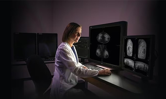
ABUS provides a “3-D” image of the breast using ultrasound technology, proven by research to be particularly useful in women with dense breasts.
Breast cancer is the second most common cancer in the world. Among women, it is the most common cause of cancer death and is by far the most common non-skin cancer. Additionally, breast cancer accounts for 25% of all new female cancer diagnoses each year and for 15% of all cancer deaths in women each year.
Given these startling facts, how do we begin to beat this dreadful disease?
Research indicates that early diagnosis through screening mammography has dramatically impacted the survival rates of women with breast cancer. Mammography is a low dose X-ray of breast tissue and is performed to detect early signs of breast cancer (preferably before the tumor is large enough to be detected on physical exam).
When a cancer can be detected early, the survival rate has been shown to be dramatically improved. The majority of mammography performed at the NMMC Breast Care Center now employs tomosynthesis/3-D technology in which multiple images of each breast are performed in an arc across the breast tissue and then computerized images are provided allowing significantly better detail of the breast tissue and of any possible developing abnormalities.
The American College of Radiology, The American Society of Breast Surgeons, the National Comprehensive Cancer Network and the Society of Breast Imagers recommend beginning annual screening mammography at the age of 40 for women of average risk.
Unfortunately, while mammography is essential to detect certain common types of breast cancer, screening mammography is not a perfect tool. Since the breast is composed of a mixture of fibrous and glandular tissue, along with interspersed fat tissue, there are portions of the breast that are more difficult to fully evaluate.
Mammography relies on X-rays to penetrate the tissue and on compression of the breast to disperse dense tissue. Because of this, mammography is limited in patients with more dense, and therefore less compressible, tissue.
In women with less dense tissue, the sensitivity of mammography (or ability to detect an abnormality) is about 90%, meaning rarely will a developing cancer go undetected with mammography. However, in women with more dense breast tissue, the sensitivity is decreased considerably, as low as 30% in those with very dense breast tissue.
So how do you determine your particular breast density?
A patient’s breast density is determined by the radiologist upon interpretation of your mammogram and is based on the relative distribution of fibroglandular tissue to fatty tissue throughout each breast. A category of density is assigned to each mammogram and the patient subsequently will be informed of their density via the mammography report and separately through written communication.
There are four potential categories of breast density: Class A, almost entirely fatty breast
- Class B, scattered areas of fibroglandular density
- Class C, heterogeneously dense breast
- Class D, extremely dense breast.
Classes A and B are considered “non-dense” and Classes C and D are considered “dense.”
Does breast density matter?
It is important to be aware of your density because increased breast density is a known risk factor for the development of breast cancer. Additionally, those patients with dense breast tissue have been shown to benefit from additional screening techniques. These include breast MRI, breast ultrasound, contrast-enhanced mammography and radionuclide breast imaging.
One of the more promising of these recently has been implemented at NMMC Breast Care Center. This painless technology, Automated Whole Breast Ultrasound (ABUS), is offered primarily to women with very dense breasts.
ABUS provides a “3-D” image of the breast using ultrasound technology, proven by research to be particularly useful in women with dense breasts. There is no associated radiation and no intravenous contrast is required. This FDA-approved, relatively low-cost technology is not user dependent and is reproducible year after year, which allows your radiologist to compare annual studies and potentially detect subtle developing changes within the breast. When combined with screening mammography, ABUS has been shown to improve cancer detection rates by at least 30%. We are excited to offer this new technology in our attempts to improve early cancer detection in our patients who are at increased risk, either due to dense breast parenchyma, family history, genetic predisposition or other lifestyle factors.
Talk with your provider about your breast density, your breast cancer risks, and your annual mammography screening schedule. And, if you are one of our many patients who have been shown to have dense breast parenchyma, let us help you schedule your screening ABUS exam today.
Mary Linda Moss, MD
Radiology
Dr. Moss is a breast imaging radiologist working with the Breast Care Center on the North Mississippi Medical Center campus in Tupelo. She joined NMMC in 2018. She graduated summa cum laude with a bachelor’s degree from Belhaven College in Jackson in 1988. She completed her medical degree cum laude from the University of Mississippi School of Medicine in Jackson, where she also completed her diagnostic and interventional radiology residency. She completed a master’s degree and fellowship in Anti-Aging and Regenerative Medicine at the University of Florida. She is board certified by the American Board of Radiology.
MyChart (also known as myConnection)
Schedule your next appointment, start an E-visit, communicate with your doctor, request prescription refills and access your test results — just a click away.
Related Links
Topics
- Managing Your Health
Share
Schedule a mammogram online or call Centralized Scheduling at (662) 377-6655 or 1-866-912-1486.

Subscribe to Our Newsletter
Like this content and want to get more? Sign up for True North, the health and wellness newsletter from North Mississippi Health Services!
Subscribe to Our Newsletter
Like this content and want to get more? Sign up for True North, the health and wellness newsletter from North Mississippi Health Services!

Nurse Link®
Not sure if you need Urgent Care or the ER? Call 1-800-882-6274 anytime to speak directly to a registered nurse and get immediate answers. Using computerized medical protocols, nurses direct callers to the most appropriate treatment. Our nurses are available 24 hours per day, seven days per week.